Diagram Of Hip.and Back.muscles - Anatomy Of Muscles Hip And Lower Back See More Anatomy Of Muscles Hip And Lower Back Anatomy And Back Hip Lower Muscle Anatomy Human Body Anatomy Body Anatomy - It joins the lower limb to the pelvic girdle.
bymagaduchane-
0
Diagram Of Hip.and Back.muscles - Anatomy Of Muscles Hip And Lower Back See More Anatomy Of Muscles Hip And Lower Back Anatomy And Back Hip Lower Muscle Anatomy Human Body Anatomy Body Anatomy - It joins the lower limb to the pelvic girdle.. · the iliopsoas muscle, which. Daniel nelson on january 1, 2019 2 comments 🔥! Topographically the muscles in this group are classed along with the lateral torso in broad terms the extrinsic muscles of the back are innervated by the ventral branches of the spinal nerves and individual cranial nerves. The quick answer to this question is the muscles of the lower back are the multifidus, longissimus, spinalis, and quadratus lumborum. Extension, flexion, adduction, and abduction.
This article will introduce the muscles in each group and touch on their origin, insertion, function, and innervation. The quads make up about 70% of the thigh's muscle mass. The muscles of the lower back help stabilize, rotate, flex, and extend the spinal column, which is a bony tower of 24 vertebrae that gives the body structure and houses the spinal cord.the spinal. The main muscles of the hip and pelvis consistsof the iliopsoas, pectinues, rectus femoris and sartorius at the front. The anatomy of your back muscles can be complex.
Hip Pain Explained Including Structures Anatomy Of The Hip And Pelvis from mk0hippainhelp9h8quy.kinstacdn.com The anatomy of your back muscles can be complex. The main action of the adductors is to pull the leg inward toward the other leg. The hip muscles encompass many muscles of the hip and thigh whose main function is to act on the thigh at the hip joint and stabilize the pelvis.without them, walking would be impossible. Make sure you're stretching to the point of tension, not pain; Hold each of these lower back and hip stretches for at least 15 to 30 seconds, and repeat several times on each side. Hip flexion is maximal with a high, forward kick that brings the leg above the level of the waist. The main muscles of the hip and pelvis consistsof the iliopsoas, pectinues, rectus femoris and sartorius at the front. Gluteal muscles, located on the back of the hip (buttocks) · the adductor muscles on the inner thigh bring.
Aarp fitness ambassador denise austin walks you through three easy stretches for hip pain.
L2, l3, and l4 spinal nerves provide sensation to the front part of your thigh and along the inner side of your lower leg. They can be divided into three main groups: The hip abductors consist of the. Vastus medialis, intermedius, lateralis and rectus femoris muscles. Extension, flexion, adduction, and abduction. The hip muscle diagram below shows a number of the muscles we will be discussing in the next sections. Creatine is now proving to be one of the most potent muscle growth accelerators giving excellent muscle mass increase and phenomenal strength increases order yours today. The muscles of the anterior thigh consist of the quadriceps (or quads): The gluteus maximus is rather large, and makes up the most prominent area of the buttocks. Lie on your back, knees bent, feet flat on the floor. The gluteals make up the muscles of the buttocks on the back of the hip. This stretch targets the muscles across the front of your hips. Raise your hips to form a straight line from your shoulders to your knees (use a support if needed).
It is a deep muscle that originates from the lower back and pelvis, and extends up to the inside surface of the upper part of the. Gluteal muscles, located on the back of the hip (buttocks) · the adductor muscles on the inner thigh bring. The hip muscles encompass many muscles of the hip and thigh whose main function is to act on the thigh at the hip joint and stabilize the pelvis.without them, walking would be impossible. The four groups are the anterior group, the posterior group, adductor group, and finally the abductor group. The anterior muscle group features muscles.
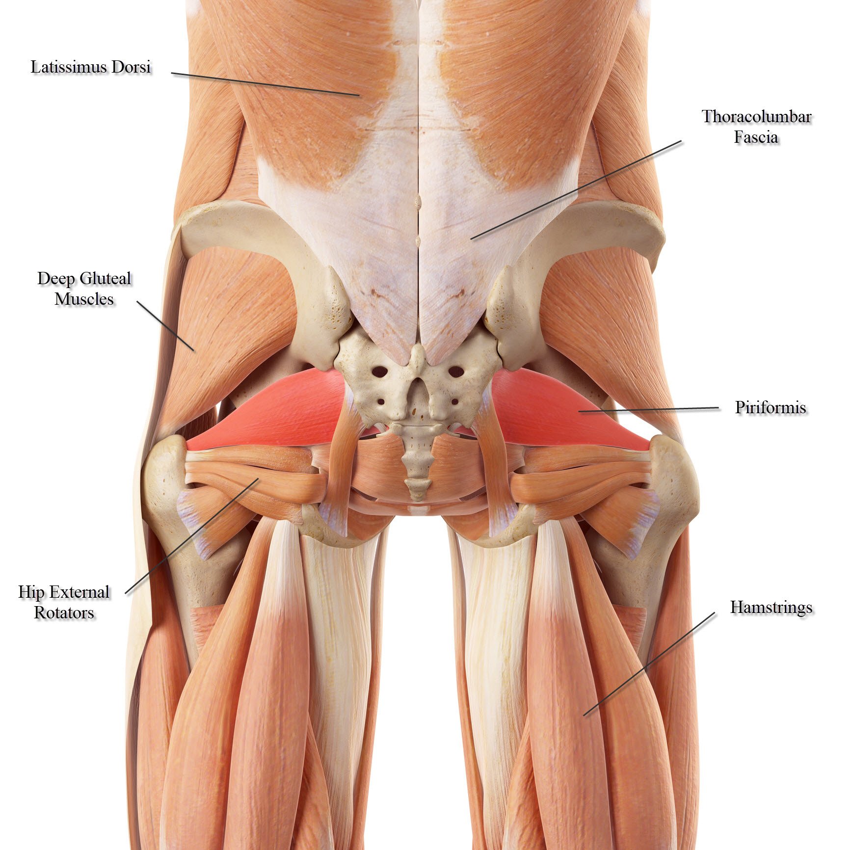
Lower Back Muscle Anatomy And Low Back Pain from ix-cdn.b2e5.com The muscles you probably know the best are your glutes (gluteal muscles), the large, strong muscles that attach to the back of your hip bones and comprise the buttocks. Topographically the muscles in this group are classed along with the lateral torso in broad terms the extrinsic muscles of the back are innervated by the ventral branches of the spinal nerves and individual cranial nerves. Creatine is now proving to be one of the most potent muscle growth accelerators giving excellent muscle mass increase and phenomenal strength increases order yours today. To learn more about the anatomy of the spine, watch this video. The muscles of the hip can be divided into three different groups. This muscle starts from the inner surface of the pelvis in the saddle region, then runs out through the back of the pelvis and across the back of the hip, through the lower buttock region (figure 2.15). It joins the lower limb to the pelvic girdle. The four groups are the anterior group, the posterior group, adductor group, and finally the abductor group.
The muscles of the hip can be divided into three different groups.
Anterior muscles (front), posterior (back), and medial (inside). Gluteal muscles, located on the back of the hip (buttocks) · the adductor muscles on the inner thigh bring. The main muscles of the hip and pelvis consistsof the iliopsoas, pectinues, rectus femoris and sartorius at the front. This muscle starts from the inner surface of the pelvis in the saddle region, then runs out through the back of the pelvis and across the back of the hip, through the lower buttock region (figure 2.15). Lower back muscle diagram anatomy does degenerative disc disease affect the lower back muscle? The vertebral column of the lower back includes the five lumbar vertebrae, the sacrum, and the coccyx. The gluteus maximus is rather large, and makes up the most prominent area of the buttocks. Some of the links in the post above are affiliate links.. The back's muscles start at the top of the back (named the cervical vertebrae) and go to the tailbone (also named the coccyx). The muscles that flex the hip are in front of the hip joint. The quads make up about 70% of the thigh's muscle mass. The bones of the pelvis and lower back work together to support the body's weight, anchor the abdominal and hip muscles, and protect the delicate vital organs of the vertebral and abdominopelvic cavities. The hip muscle diagram below shows a number of the muscles we will be discussing in the next sections.
The muscles you probably know the best are your glutes (gluteal muscles), the large, strong muscles that attach to the back of your hip bones and comprise the buttocks. The human back extends from the buttocks to the posterior portion of the neck and shoulders. This is a diagram of the larger and more surface muscles of the low back. Gluteal muscles, located on the back of the hip (buttocks) · the adductor muscles on the inner thigh bring. L2, l3, and l4 spinal nerves provide sensation to the front part of your thigh and along the inner side of your lower leg.
Low Back Pain Anything But A Dream For Rowers from sportsinjury.wpengine.com To put it plainly, sometimes hip pain comes from the hip, but a lot of times hip pain comes from the back. When these muscles become tight due to inadequate activity (such as from a sedentary lifestyle), they become shorter, and in turn, cause tension around the sacroiliac. It is opposite from the chest, and the vertebral column runs down the back. Daniel nelson on january 1, 2019 2 comments 🔥! Gastrocnemius gastrocnemius muscle, large posterior muscle of the calf of the leg. Make sure you're stretching to the point of tension, not pain; The hip muscle diagram below shows a number of the muscles we will be discussing in the next sections. Vastus medialis, intermedius, lateralis and rectus femoris muscles.
One of the adductor muscles of the hip flexor, its main function is to adduct the thigh.
The inner thigh is formed by the adductor muscles. To put it plainly, sometimes hip pain comes from the hip, but a lot of times hip pain comes from the back. L2, l3, and l4 spinal nerves provide sensation to the front part of your thigh and along the inner side of your lower leg. Hold each of these lower back and hip stretches for at least 15 to 30 seconds, and repeat several times on each side. Study flashcards on chapter 10 muscle diagrams at cram.com. · the iliopsoas muscle, which. Another common cause of lower back and hip pain is disc injury. Make sure you're stretching to the point of tension, not pain; The hip is surrounded by thick muscles. There are several different layers of muscles in your back that are often pulling in different and various directions. The muscles of the lower back help stabilize, rotate, flex, and extend the spinal column, which is a bony tower of 24 vertebrae that gives the body structure and houses the spinal cord.the spinal. The muscles you probably know the best are your glutes (gluteal muscles), the large, strong muscles that attach to the back of your hip bones and comprise the buttocks. The diagram shows the posterior (rear) view of the buttock.